The William C. Davis Monoclonal Antibody Center
It is with great sadness that we share the passing of Dr. Davis. While this is a significant loss for our team and community, we remain committed to honoring his vision by continuing the important work of the service center.
As we navigate this transition, some services and response times may be slower than usual. We appreciate your patience and understanding as we adapt and move forward.
We are grateful for your continued support and look forward to serving you for many years to come.
If you have questions or concerns, please contact us.
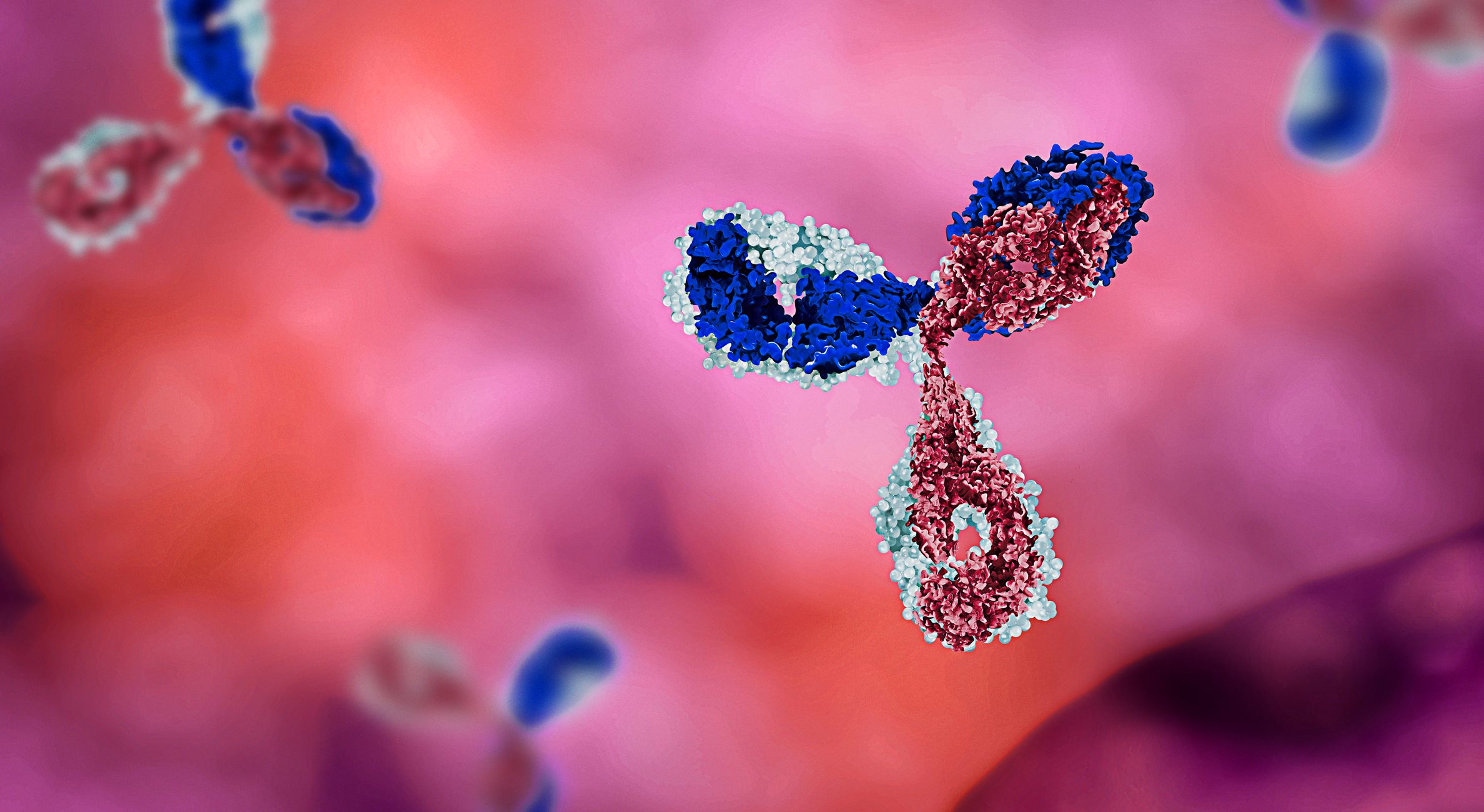
The Monoclonal Antibody Center provides a range of services related to the production and characterization of monoclonal antibodies to researchers and clinicians affiliated with WSU.
Monoclonal antibodies to leukocyte differentiation molecules, activation molecules, and MHC Class I and II molecules in many species of food and companion animals have been produced by the WSU Monoclonal Antibody Center. All antibodies are developed in mice.
Taxonomic Key Program is a searchable database that allows researchers to easily identify monoclonal antibody-defined molecules by their physical characteristics.
Isotype controls
| Catalog # | mAb | Ig isotype | E coli J5 |
|---|---|---|---|
| COL2001 | COLIS69A | IgG1 | E coli J5 |
| COL2002 | COLIS205C | IgG2a | E coli J5 |
| COL2003 | COLIS169A | IgG2b | E coli J5 |
| COL2004 | COLIS41B | IgG3 | E coli J5 |
| COL2005 | COLIS52A2 | IgM | E coli J5 |
Monoclonal antibodies by species
| Clone | Ig Isotype | Specificity | Bison | Llama and alpaca | Cat | Cattle | Chicken | Dog | Dolphin | Ferret | Goat | Hamster | Horse | Pig | Rabbit | Sheep | Water Buffalo |
| 8000000 | G1 | CD3 | X | ||||||||||||||
| 17D | G1 | CD4 | X | X | |||||||||||||
| 209MD26A | G2a | CD209 | X | X | |||||||||||||
| 25-2A | G1 | CD8 | X | ||||||||||||||
| 25-2C (D422) | M | Predicted specificity CD45 | X | ||||||||||||||
| 73/6.9.1 | G3 | CD8α | X | ||||||||||||||
| 74-11-10 | Gb2 | MHC I | X | ||||||||||||||
| 74-12-4 | G2b | CD4 | X | ||||||||||||||
| 74-22-15 | G1 | CD172a (SWC3) | X | ||||||||||||||
| 74-22-15A | G2b | CD172a (SWC3) | X | ||||||||||||||
| 74-9-3A1 | M | CD45 | X | ||||||||||||||
| 76-2-11 | G2a | CD8α | X | ||||||||||||||
| 76-7-4 | G2a | B cell and thymocytes | X | ||||||||||||||
| 7C2B | G2a | CD8α | X | X | X | X | X | ||||||||||
| 86D | G1 | TCR1-N6 γ chan | X | X | |||||||||||||
| 86D1 | G1 | TCR1-N7 cluster, γ chain specific? | X | ||||||||||||||
| 8E6-2b3c | G2b | CD3 | X | ||||||||||||||
| 8E6-8C8 | G2a | CD3 | X | ||||||||||||||
| AC306B | G1 | Granulocyte monocyte | X | ||||||||||||||
| AV114A | M | Thrombocytes | X | ||||||||||||||
| AV129A | M | Pan leukocyte | X | ||||||||||||||
| AV14A | M | Pan leukocyte | X | ||||||||||||||
| AV18A | M | Pan leukocyte | X | ||||||||||||||
| AV191A | M | Late B | X | ||||||||||||||
| AV213A | G2a | CD8 | X | ||||||||||||||
| AV243A | G2a | Granulocyte monocyte | X | ||||||||||||||
| AV261C | G3 | PanT | X | ||||||||||||||
| AV309A | G1 | CD4 | X | ||||||||||||||
| AV319A | M | CD8 | X | ||||||||||||||
| AV36A | M | Pan leukocyte | X | ||||||||||||||
| AV43A | G3 | Pan leukocyte | X | ||||||||||||||
| AV47A | sIgM | sIgM | X | ||||||||||||||
| AV64A | G1 | MHC II | X | ||||||||||||||
| AV66D | G3 | CD4 | X | ||||||||||||||
| AV66D | G3 | CD4 | X | ||||||||||||||
| AV67A | G1 | αβ T | X | ||||||||||||||
| AV92A | M | CD4 | X | ||||||||||||||
| B18A | G1orG3 | CD9 | X | X | |||||||||||||
| B29A | G2a | CD5 | X | X | |||||||||||||
| B5C | G2b | MHC I | X | ||||||||||||||
| B7A | M | WC1+ γδ T cell N1 epitope | X | X | |||||||||||||
| B7A1 | M | WC1-N1 | X | X | X | X | X | ||||||||||
| BAG25A | M | WC1+ γδ T cell N28 epitope | X | ||||||||||||||
| BAG2B | M | WC1+ γδ T cell N27 epitope | X | ||||||||||||||
| BAG40A | G3 | CD44 | X | X | X | X | X | X | X | X | X | ||||||
| BAG40A | M- G3 | CD44 | X | M | |||||||||||||
| BAGB20A | G1 | CD45 predicted | X | ||||||||||||||
| BAGB45A | G1 | CD5 | X | ||||||||||||||
| BAQ111A | M | CD8 | X | X | X | ||||||||||||
| BAQ125A | G2b | Platelets | X | ||||||||||||||
| BAQ128A | G1 | WC1+ γδ T cell | X | ||||||||||||||
| BAQ151A | G1 | CD11c | X | ||||||||||||||
| BAQ153A | M | CD11c | X | X | X | X | X | ||||||||||
| BAQ155A | G1 | B cells | X | X | |||||||||||||
| BAQ159A | G1 | WC1+ γδ T cell N4 subset | X | X | |||||||||||||
| BAQ15A | M | CD21 | X | X | |||||||||||||
| BAQ30A | G1 | CD18 | X | X | X | X | X | X | X | X | X | X | X | X | |||
| BAQ44A | M | B cells | X | X | X | X | |||||||||||
| BAQ4A | G1 | WC1-N2 | X | X | X | X | X | ||||||||||
| BAQ56A | G1 | Platelets | X | ||||||||||||||
| BAQ83A | G2b | CD6 | X | X | |||||||||||||
| BAQ89A | G1 | WC1-N4 | X | X | X | X | X | ||||||||||
| BAQ91A | G1 | CD6 | X | X | X | ||||||||||||
| BAQ92A | G1 | CD62L | X | X | X | ||||||||||||
| BAQ95A | G1 | CD2 | X | X | X | X | |||||||||||
| BAT31A | G1 | CD44 | X | X | X | X | X | X | |||||||||
| BAT72A | G1 | CD8β | X | ||||||||||||||
| BAT82A | G1 | CD8β | X | X | X | X | |||||||||||
| BIB | M | ab and gd T cells | X | ||||||||||||||
| BIG312D3 | G1 | IgA | X | ||||||||||||||
| BIG501E | G1 | λ light chain | X | X | X | X | X | ||||||||||
| BIG715A | G1 | IgG1 | X | X | X | X | |||||||||||
| BIG73A | G1 | sIgM | X | X | X | ||||||||||||
| Bo-IL-22 | G1 | IL-22 | X | X | |||||||||||||
| CACT105A | G1 | CD5 | X | ||||||||||||||
| CACT108A | G2a | CD25 | X | X | |||||||||||||
| CACT114A | G2b | CD26 | X | ||||||||||||||
| CACT116A | G1 | CD25 | X | X | X | X | X | ||||||||||
| CACT130A | G3 | CD8α | X | ||||||||||||||
| CACT138A | G1 | CD4 | X | X | X | ||||||||||||
| CACT152A | M | ACT31 | X | ||||||||||||||
| CACT153A | M | ACT37 | X | X | |||||||||||||
| CACT180A | G1 | ACT34 | X | X | X | ||||||||||||
| CACT191A | M | ACT32 | X | ||||||||||||||
| CACT195A | M | ACT27 | X | ||||||||||||||
| CACT200A | G1 | ACT1 | X | X | |||||||||||||
| CACT200A | G1 | SLAMF9 | X | ||||||||||||||
| CACT206A | G2a | ACT1 | X | X | |||||||||||||
| CACT206A | G2a | SLAMF9 | X | ||||||||||||||
| CACT216A | M | CD50 - ACT28 | X | X | X | ||||||||||||
| CACT21A | G1 | WC1+ γδ T cell N31 epitope | X | ||||||||||||||
| CACT225A | G1 | ACT29 | X | ||||||||||||||
| CACT22B | G1 | TCR1-N7 γ chain specific? | X | ||||||||||||||
| CACT243A | M | ACT38 | X | ||||||||||||||
| CACT26A | G1 | ACT2 | X | X | |||||||||||||
| CACT26A | G2b | CD26 | X | ||||||||||||||
| CACT282A | M | ACT35 | X | ||||||||||||||
| CACT31A | CD2 | M | X | ||||||||||||||
| CACT61A | M | TCR1 δ chain specific | X | ||||||||||||||
| CACT7A | M | ACT1 | X | ||||||||||||||
| CACT80C | G1 | CD8α | X | X | X | X | X | ||||||||||
| CACT83B | M | CD4 | X | ||||||||||||||
| CACT83B | M | CD4 | X | ||||||||||||||
| CACTB21A | G1 | WC1+ γδ T cell N29 | X | ||||||||||||||
| CACTB31A | G2b | WC1+ γδ T cell N30 | X | ||||||||||||||
| CACTB32A | G1 | WC1-N3 | X | X | X | X | |||||||||||
| CACTB51A | G2a | CD45 | X | ||||||||||||||
| CACTB6A | M | TCR1-N6 γ chain specific? | X | X | X | X | |||||||||||
| CACTB7A | G1 | WC1+ γδ T cell N3epitope | X | X | X | ||||||||||||
| CACTB81A | G1 | TCR1-N7 γ chain specific? | X | ||||||||||||||
| CADO13A | M | Neutrophils | X | ||||||||||||||
| CADO18A | G1 | Pan leukocyte | X | ||||||||||||||
| CADO19A | G1 | CD45 predicted | X | ||||||||||||||
| CADO21A | G2b | CD47 predicted | X | ||||||||||||||
| CADO46A | G1 | CD8 | X | ||||||||||||||
| CADO48A | G1 | Neutrophils | X | ||||||||||||||
| CADO6A | G1 | X | |||||||||||||||
| CAM36A | G1 | CD14 LPS receptor (monocytes, variable on granulocytes in different species) | X | X | X | X | X | X | X | X | X | X | |||||
| CAM66A | M | CD14 LPS receptor (monocytes, variable on granulocytes in different species) | X | X | X | X | X | X | |||||||||
| CAPP2A | G1 | CD41 | X | X | X | X | X | ||||||||||
| CAT30A | G1 | CD4 | X | ||||||||||||||
| CAT82A | G1 | MHC II | X | X | |||||||||||||
| CF26A | G1 | Pan lymphocyte | X | ||||||||||||||
| CF54A | G1 | Pan T1 | X | ||||||||||||||
| CF74A | G1 | Pan T2 | X | ||||||||||||||
| CF77A | G1 | Pan lymphocytes and monocytes | X | ||||||||||||||
| CF7A | G1 | T sub | X | ||||||||||||||
| CH138A | M | Granulocytes | X | X | X | ||||||||||||
| CL2A | G1 | CD41 Platelets | X | X | X | ||||||||||||
| CL35A | G1 | Neutrophil | X | ||||||||||||||
| CT1.572 (T572) | G2a | CD5 | X | ||||||||||||||
| CT3.357 (T357) | M | CD8β | X | ||||||||||||||
| CTOKT8 | G2a | CD8 | X | ||||||||||||||
| DH13A | M | CD5 | X | ||||||||||||||
| DH16A | M | CD45RB | X | X | X | X | |||||||||||
| DH24A | M | CD90 - granulocytes | X | X | |||||||||||||
| DH29A | M | CD4 | X | ||||||||||||||
| DH2A | M | CD90 | X | ||||||||||||||
| DH3B | G1 | CD5 | X | ||||||||||||||
| DH59B | G1 | CD172a | X | X | X | X | X | X | X | X | X | ||||||
| DLL132A | M | CD21 | X | ||||||||||||||
| DLL20A | M | B and granulocytes | X | ||||||||||||||
| DLL46A | G1 | X | |||||||||||||||
| DLL51A | G1 | B | X | ||||||||||||||
| DLL76A | G1 | B | X | ||||||||||||||
| DOL10 | IL10 | X | X | ||||||||||||||
| Do-TNF-α | G1 | TNF-α | X | X | X | ||||||||||||
| DU1-29 | G1 | CD62L | X | X | |||||||||||||
| E38A | G1 | CD45R0 predicted | X | ||||||||||||||
| EC1.1 | G1 | CD335 | X | ||||||||||||||
| ETC142B1A | G2a | CD8α | X | ||||||||||||||
| ETC142B1A | G2a | CD8α | X | ||||||||||||||
| F46A | G1 | B cell | X | X | X | ||||||||||||
| F72A | G1 | Granulocytes, monocytes, B | X | ||||||||||||||
| FOX20A | G2b | FoxP3 | X | ||||||||||||||
| FOX5A | G1 | Foxp3 | X | X | |||||||||||||
| FW3-218 | G2b | CD49d | X | X | X | X | |||||||||||
| FW4-101 | G1 | CD29 | X | X | X | X | X | ||||||||||
| GB110A | M | ACT16 | X | X | |||||||||||||
| GB112A | G1 | CD25 | X | X | X | ||||||||||||
| GB21A | G2b | TCR1-N24 δ chain specific | X | X | X | ||||||||||||
| GB25A | G1 | CD21 | X | X | X | X | |||||||||||
| GB26A | M | B cells | X | X | |||||||||||||
| GB45A | G1 | WC1-N26 | X | X | X | X | |||||||||||
| GB50A | G1 | Pan leukocyte, endothelial cells | X | ||||||||||||||
| GB53A | G1 | Gr, mono/ lymph sub | X | ||||||||||||||
| GB54A | G2a | WC1+ γδ T cell N25 epitope | X | X | X | ||||||||||||
| GB54A | G2a | WC1-N25 | X | ||||||||||||||
| GC1A | G2a | CD4 | X | X | |||||||||||||
| GC34A | M | Lymphocytes, monocytes | X | ||||||||||||||
| GC42A | G1 | CD45R0 | X | X | |||||||||||||
| GC50A | M | CD4 | X | X | X | X | X | ||||||||||
| GC5A | G1 | Platelets | X | ||||||||||||||
| GC6A | M | CD45R | X | X | X | X | |||||||||||
| GRAN2A1 | G1 | Granulysin | X | ||||||||||||||
| GS23A | G1 | Neutrophils | X | ||||||||||||||
| GS5A | G1 | CD45R | X | X | X | X | |||||||||||
| H1A | G2a | MHC I | X | ||||||||||||||
| H20A | G1 | CD18 | X | ||||||||||||||
| H22A | G2a | CD44 | ? | X | X | ||||||||||||
| H34A | G2b | MHC II | X | ||||||||||||||
| H42A | G2a | MHC II | X | X | X | X | X | X | X | X | |||||||
| H58A | G2a | MHC I | X | X | X | X | X | X | X | X | X | X | X | ||||
| HAB1A | G1 | CD4 | X | ||||||||||||||
| HAB2A | G1 | Pan T | X | ||||||||||||||
| HAB6B = HASA18A | G2a | Pan lymphocyte | X | ||||||||||||||
| HACT39A | G1 | Granulocytes | X | ||||||||||||||
| HAL16A | G1 | MHC II | X | ||||||||||||||
| HAL17A | G2a | B | X | ||||||||||||||
| HAL36A | G2a | CD4 | X | ||||||||||||||
| HASA18A | M | Pan lymphocyte | X | ||||||||||||||
| HASA25A | G1 | CD45 Predicted | X | ||||||||||||||
| HASA26B | G1 | Granulocytes | X | ||||||||||||||
| HASA7A | G1 | B | X | ||||||||||||||
| HAT13A | G2b | CD45 Predicted | X | ||||||||||||||
| HAT19A | G2a | Pan T = HAB2A | X | ||||||||||||||
| HB19A | G2a | CD5 | X | ||||||||||||||
| HB32A | G2a | CD45R0 predicted | X | ||||||||||||||
| HB61A | G1 | CD4 | X | ||||||||||||||
| HB65A | G2a | CD4 sub | X | ||||||||||||||
| HB72A | G2a | CD45R0 predicted | X | ||||||||||||||
| HB86A | G1 | CD4 sub | X | ||||||||||||||
| HB88A | G1 | CD2 | X | ||||||||||||||
| HT14A | G1 | CD8β | X | ||||||||||||||
| HT23A | G1 | CD5 | X | ||||||||||||||
| HUH71A | G1 | CD47 | X | X | X | ||||||||||||
| HUH73A | G1 | CD11a | X | X | X | X | X | X | X | X | |||||||
| HUH82A | G2a | CD18 | X | X | X | X | X | X | X | ||||||||
| I LA158A | G1 | CD40 | X | ||||||||||||||
| IL11A | G2a | CD4 | X | X | |||||||||||||
| IL-17A | G1 | Bo-IL-17A | X | X | |||||||||||||
| ILA114A | G1 | CD205 | X | ||||||||||||||
| ILA116A | G3 | CD45R0 | X | X | X | X | X | ||||||||||
| ILA-11A | G2a | CD4 | X | X | |||||||||||||
| ILA158A | G1 | CD40 | X | X | |||||||||||||
| ILA159 | G1 | CD80 | X | X | |||||||||||||
| ILA190A | G1 | CD86 | X | X | |||||||||||||
| ILA29 | G1 | WC1+ γδ T cell | X | X | |||||||||||||
| ILA29A | G1 | WC1 | X | ||||||||||||||
| ILA53A | G2a | CD205 | X | ||||||||||||||
| ILA77A | M | CD71 | X | X | |||||||||||||
| ISC16A | M | CD8 | X | ||||||||||||||
| ISC18A | G2a | CD45 | X | ||||||||||||||
| ISC27A | G2a | CD8 | X | ||||||||||||||
| ISC29E | G1 | CD8 | X | ||||||||||||||
| ISC38A | G1 | CD8 | X | ||||||||||||||
| ISC39A | G1 | CD45 | X | ||||||||||||||
| ISC76A | M | CD45 | X | ||||||||||||||
| KTSN7A | G1 | CD69 | X | X | X | ||||||||||||
| LCT27A | G1 | CD45R | X | ||||||||||||||
| LCTB2A | G3 | CD25 | X | X | X | X | X | ||||||||||
| LCTB32A | M | CD25 | X | X | |||||||||||||
| LCTB6A | G1 | ACT9 | X | ||||||||||||||
| LH41A | M | B | X | ||||||||||||||
| LH43A | G1 | CD2/CD6 like | X | ||||||||||||||
| LH7A | M | B sub | X | ||||||||||||||
| LH9A | M | ACT1 | X | ||||||||||||||
| LND37A | G1 | Monocytes | X | ||||||||||||||
| LND41A | G1 | DC and a subset of innate lymphoid cells | X | ||||||||||||||
| LND68A | G1 | Monocytes | X | X | X | ||||||||||||
| LT10A | G2a | CD2/CD6 like | X | ||||||||||||||
| LT12A | G2a | CD45 predicted | X | ||||||||||||||
| LT1A | G2a | MHC II | X | ||||||||||||||
| LT36A | G2b | CD44 | X | X | |||||||||||||
| LT3A | G1 | CD5 like | X | ||||||||||||||
| LT41A | G2a | CD44 | X | X | |||||||||||||
| LT48A | G2a | CD11a | X | ||||||||||||||
| LT4A | G2a | CD5 like | X | ||||||||||||||
| LT5A | G2a | CD8 | X | ||||||||||||||
| LT78A | M | B sub | X | ||||||||||||||
| LT79A | G3 | B sub | X | ||||||||||||||
| LT86A | G2a | CD9 | X | X | |||||||||||||
| LT97A | G2b | Pan T | X | ||||||||||||||
| MM10A | G2b | CD11b | X | X | |||||||||||||
| MM12A | G1 | CD11b | X | X | X | X | X | ||||||||||
| MM1A | G1 | CD3 | X | X | X | ||||||||||||
| MM20A | G1 | Granulocytes = CH138A | X | X | |||||||||||||
| MM20A | G1 | Neutrophils/monocytes | X | ||||||||||||||
| MM61A | G1 | CD14 | X | ||||||||||||||
| MRB102A | M | Pan lymphocyte | X | ||||||||||||||
| MRB107A | G1 | B sub | X | ||||||||||||||
| MRB120A | G1 | Granulocytes, basophils, monocytes | X | ||||||||||||||
| MRB25A | M | B | X | ||||||||||||||
| MRB61A | G1 | T3 | X | ||||||||||||||
| MSA3 | G2a | MHC II HLA DR orthologue | X | ||||||||||||||
| MSA4 | G2a | CD2 | X | ||||||||||||||
| MUC127A | G2a | TCR1 γ chain | X | ||||||||||||||
| MUC2A | G2a | CD2 | X | X | |||||||||||||
| MUC2A | G2a | CD2 | X | X | X | X | |||||||||||
| MUC76A | G2a | CD11a | X | ||||||||||||||
| MUC93A | G1 | CD59 need validation | X | ||||||||||||||
| NK29A | G1 | CD26 | X | X | |||||||||||||
| NK29A | G1 | NK Cluster 2 activation molecule | X | ||||||||||||||
| NK47A | G2a | CD26 | X | ||||||||||||||
| NK47A | G2a | NK Cluster 2 | X | ||||||||||||||
| NK64A | G1 | Gp96 | X | X | |||||||||||||
| NK64A | G1 | NK Cluster 1 | X | ||||||||||||||
| OKT8 | G2a | CD8 | X | ||||||||||||||
| PG104A | M | CD8+ CD4 sub, γδ small sub | X | ||||||||||||||
| PG106A | M | Pan lymphocyte | X | ||||||||||||||
| PG114A | G1 | CD5 | X | ||||||||||||||
| PG124A | G1 | CD27 (SWC2) | X | ||||||||||||||
| PG145A | M | sIgM | X | ||||||||||||||
| PG164A | G2a | CD8β | X | ||||||||||||||
| PG167A | G1 | CD45RA | X | ||||||||||||||
| PG168A | G3 | CD2 | X | ||||||||||||||
| PG68A | G1 | Granulocytes | X | ||||||||||||||
| PG74C | M | Subsets γδ CD4 CD8 T cells | X | ||||||||||||||
| PG77A | M | CD45RA | X | ||||||||||||||
| PG83A | G3 | SWC4 | X | ||||||||||||||
| PG89A | G1 | Pan lymphocyte | X | ||||||||||||||
| PG90A | G1 | CD6 | X | ||||||||||||||
| PG92A | M | SWC5 | X | ||||||||||||||
| PG94A | M | Subset SWC6+ γδ T cells | X | ||||||||||||||
| PG96A | G1 | CD45RA | X | ||||||||||||||
| PG97A | M | CD45RA | X | ||||||||||||||
| PGB78A | G2a | CD45RA | X | ||||||||||||||
| PGBL22A | G1 | TCR1 δ chain specific | X | ||||||||||||||
| PGBL23A | G1 | CD2 | X | ||||||||||||||
| PGBL24A | M | Subset γδ T cells | X | ||||||||||||||
| PGBL25A | G1 | CD25 | X | ||||||||||||||
| PGBL31A | G1 | Subset γδ T cells | X | ||||||||||||||
| PIG45A | G2b | sIgM | X | ||||||||||||||
| PIG45A2 | G2b | IgM | X | X | X | ||||||||||||
| PT14A | G2a | TCR1 γ chain | X | ||||||||||||||
| PT36A | G1 | CD8α | X | ||||||||||||||
| PT79A | G2a | TCR1 γ chaint | X | ||||||||||||||
| PT81B | G2b | CD8α | X | ||||||||||||||
| PT85A | G2a | MHC I | X | X | X | X | X | X | X | X | |||||||
| PT90A | G2a | CD4 | X | ||||||||||||||
| RACT19A | M | CD4 CD8 SUB CD28? | X | ||||||||||||||
| RACT20A | G1 | Basophils and sub CD4 CD8 | X | ||||||||||||||
| RACT38A | G1 | Pan leukocyte | X | ||||||||||||||
| RACT43A | M | Granulocytes | X | ||||||||||||||
| RACT44A | M | Granulocytes | X | ||||||||||||||
| RACT48A | G2a | CD11a | X | ||||||||||||||
| RACT48A | G2a | CD18 | X | ||||||||||||||
| RH1A | G3 | CD9 | X | X | X | X | X | ||||||||||
| RIF60A | G1 | IFN-γ | X | X | |||||||||||||
| RT18A | M | CD11b | X | ||||||||||||||
| RT19A | M | B sub | X | ||||||||||||||
| RT22A | M | T2 = RTH21A | X | ||||||||||||||
| RTH161A | G1 | CD18 | X | ||||||||||||||
| RTH186A | G1 | Pan lymphocyte | X | ||||||||||||||
| RTH192A | G1 | CD5 | X | ||||||||||||||
| RTH1A | G1 | CD4 | X | ||||||||||||||
| RTH21A | G1 | T2 | X | ||||||||||||||
| RTH26A | G2a | T4 | X | ||||||||||||||
| RTH2A | G1 | T1 | X | ||||||||||||||
| RTH32A | M | CD58 | X | ||||||||||||||
| RTH33A | G1 | CD58 | X | ||||||||||||||
| ST1A | IgG2a | CD5 | X | ||||||||||||||
| ST8 | M | CD8 | X | X | |||||||||||||
| TE1A | M | CD28 | X | X | |||||||||||||
| TH14B | G2a | MHC II HLA-DR | X | X | X | X | X | X | X | X | X | X | X | ||||
| TH16A | G2a | MHC II | X | ||||||||||||||
| TH17A | M | CD47 | X | X | X | X | X | X | |||||||||
| TH18A | G3 | Pan lymphocyte | X | ||||||||||||||
| TH21A | G2b | MHC II | X | ||||||||||||||
| TH22A | G2a | MHC II | X | ||||||||||||||
| TH81A | G2a | MHC II HLA-DQ | X | X | X | X | X | X | X | X | X | X | X | ||||
| TH97A | G2a | CD1b | X | X | |||||||||||||
| W6/32 | G2a | MHC I | X |
About us
In the mid-70s, a group of investigators demonstrated in mice that it is possible to immortalize B lymphocytes producing single monospecific antibodies (mAbs) by fusing B lymphocytes to tissue culture adapted myeloma (malignant form of an antibody producing cell) cells. The hybrid cells (hybridomas) were stable and could be maintained in culture indefinitely or preserved in liquid nitrogen for later use. These findings represented a major breakthrough in the way antibody reagents could be developed for basic and biomedical research. Mice could be immunized with antigens of interest and then used as a source of B lymphocytes to produce mAbs of known specificity. The spleen was shown to be the best source of B lymphocytes for making hybridomas. Hybridomas producing the desired mAbs could be identified by examining culture medium from primary cultures of fused cells. In subsequent investigations, myeloma fusion partners were developed that could only grow in selective growth medium if fused to a B lymphocyte, eliminating the problem of distinguishing hybridomas from cells that had not fused with a B lymphocyte. Myeloma fusion partners were later identified that could no longer produce their own antibody but could produce antibody derived from the B lymphocyte. These advances simplified and improved the methodology for producing mAbs.
Research universities, research institutes and industry established core facilities to exploit mAb technology to develop mAbs for use as analytical tools in the biological and biomedical sciences, in the characterization of the immune system, study the mechanisms by which pathogens cause disease, develop improved diagnostic assays, and identify antigens from pathogens that can be used in subunit vaccines. Because of our research needs, we started developing mAbs for use in food and companion animal research and diagnostic medicine in 1979. To encourage other investigators to use mAb technology, we sponsored the first international symposium and workshop ‘Impact of Monoclonal Antibody Technology on Animal and Plant Agricultural Research’ in June of 1982. We also participated in a workshop sponsored by the National Research Council of the National Academy of Science on ‘Priorities in Biotechnology Research for International Development held in July of 1982, a symposium sponsored by the USDA ‘Hybridoma Technology in Agricultural and Veterinary Research’ held in Oct. of 1983 and an international conference sponsored by the International Laboratory for Research on Animal Diseases ‘The Ruminant Immune System In Health and Disease’ held in Kenya Africa in September 1983. In 1984, we co-sponsored a meeting with Australia ‘Characterization of the Bovine Immune System and the Genes Regulating Expression of Immunity with Particular Reference to their Role in Disease Resistance’ held in Honolulu, Hawaii. During this period of time, we developed some of the first sets of mAbs for use in the study of the immune system in food and companion animals. We also participated in all the international workshops to characterize the first sets of mAbs.
Goals
The Washington State University Monoclonal Antibody Center was established in 1982 with the following goals:
- Provide a central facility for the production of mAbs for investigators that do not have the facilities or expertise to produce mAbs for their research.
- Develop and use mAbs to study the mechanisms regulating the immune response to pathogens and candidate subunit vaccines in veterinary species.
- Provide a source of mAbs for the research community for use in food and companion animal research.
- Provide graduate and professional training in the use of mAb technology in research and the development of diagnostic reagents.
- Provide graduate and professional training in the use of mAbs in immunology and flow cytometry.
- Collaborate with investigators requiring expertise and resources available through the center to conduct studies in immunology.
- Aid in the development of clinical flow cytometry diagnostic services in veterinary medicine.